
Art and science collide at The Brain Observatory
THE BRAIN ON ART
JANUARY-FEBRUARY 2023
The exhibition features six artists who explore how the intangible mind relates to the physical object that we call the brain.
Susan Aldworth, Andrew Carnie, Annie Cattrell, Suki Chan, Pablo Garcia Lopez, and Nina Sellars are at the forefront of an international community of artists who sustain serious interest in scientific research. They often work closely with scientists and clinicians in laboratory and healthcare settings. Several of them are based in the UK, where the creative dialogue between art and bioscience has enjoyed significant funding support since the 1990s. This art-science movement continues to contribute cultural insight and empathy to the human quest for health and well-being.
The exhibition is jointly curated by Dr. Jacopo Annese, President, and CEO of the Brain Observatory, and Dr. Marius Kwint, Reader in Visual Culture at the University of Portsmouth, UK.
Nina Sellars
SCAN
Scan (graphic wall image) laser cut adhesive-backed matte vinyl film (red and black)
Click to enlarge
Artist’s statement:
The proliferation of various anatomical images both historical and modern in science defines different approaches to the human body. These approaches are all a combination, in varying degrees, of theory, observation, culture and technology. Anatomical images are never truly objective as they are created in the context of their time, and are consequently subject to the technologies and beliefs of that time. The study of anatomy is therefore also a study of contemporary ways of thinking, seeing and managing knowledge and information.
In SCAN, the focus is on the enactment of knowledge, as it unfolds as a process, through the imaging of anatomy. Scan contains an encoded body of information in the graphic form of a QR code. The code links to, and activates, an online animation comprising MRI brain scans [of the artist]. In SCAN, the brain as an object is not only dematerialised, but also mobilised, taking it from the physical space of the gallery to the virtual space of the Internet. The brain has been encoded and decoded, and made visible through multiple acts of mediation.
Susan Aldworth
PASSING THOUGHTS
Left: Passing Thoughts 4 (archival digital print) / Right: Passing Thoughts 2 (archival digital print)
Click to enlarge
Artist’s statement:
Something happened in the studio . . . that made me realize that I could make a different type of print whilst still printing directly from the brain slice. The result is the fifteen digital photographic prints which make up the Passing Thoughts suite. I hope that they will remain mysterious. They are unaltered photographs – authentic, strange and beautiful pictures of a human brain.
They seem to capture a moment – and this we caught on camera in the studio when we were working with the brain slices. The images revealed themselves and then disappeared in seconds. They were transient and disappeared like a thought. We did not own that brain; it was lent to us. It made its mark and then it went. . . . Originally my intention was to just look at the brain as object. But the brain, in a way, turned from object to subject as we were making the work. So, they are not just anatomical works: they are about the transience of self.
Andrew Carnie
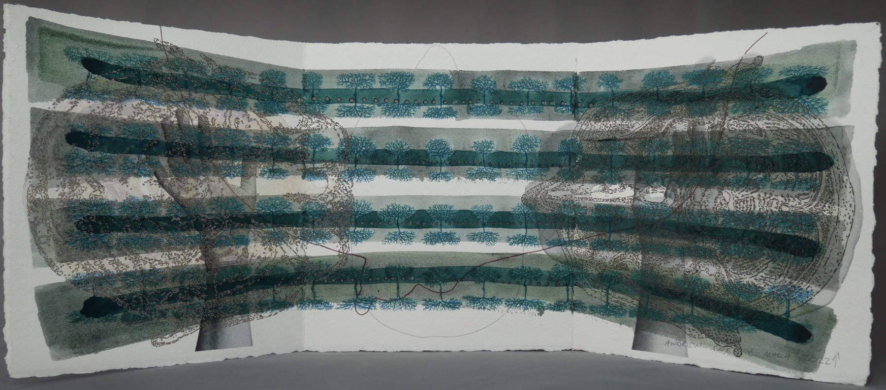
ATLAS: THERE AND HERE
Video installation, 19 minutes, 2012
Click to enlarge
Commissioned for the 2012 Wellcome Collection exhibition Brains: The Mind as Matter in London, this video is based on the Atlas of Head Sections (1893) by the pioneering Scottish surgeon William Macewen in the Wellcome Library. It reflects Carnie’s interest in the act of anatomical slicing and themes of change and mortality. Interleaving images derived from the book with more recent photographs of Macewen’s original specimens at the University of Glasgow, the video proceeds through haunting anatomical renditions of the head, occasionally returning to the more familiar domains of the medical textbook and the museum – what Carnie calls “the sphere of the living.”
CHANGE MY MIND
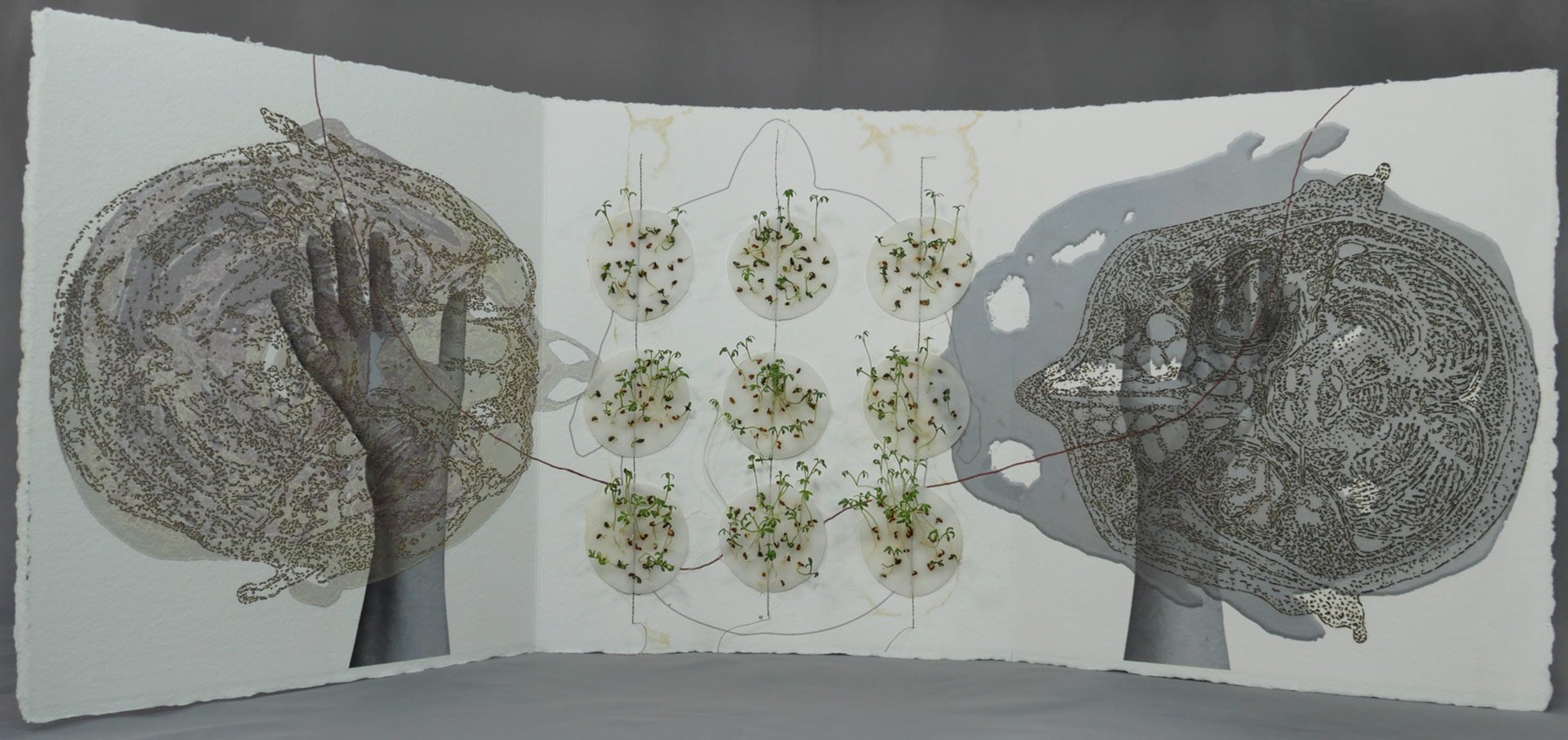

These hand-amended digital prints are from Carnie’s contribution to a multi-participant work that he initiated in response to a commission by Science Gallery Bengaluru, India, for its online exhibition and events programme Psyche. His theme was interventions in the brain through implants and other neural technologies. Other artists and scientists, mainly in the Bengaluru region, were sent basic paper prints of Carnie’s digital drawings of the brain and hands, and invited to use them to express their feelings and concerns about the mind in our technological society. Some of the remarkable creative engagements with these embryonic artworks can be seen in the online discussion event about the works.
Annie Cattrell
FROM WITHIN
Silvered bronze cast, 2006.
Click to enlarge.
Annie Cattrell is a Royal Scottish Academician and Fellow of the Royal Society of Sculptors in the UK. Her work explores poetic continuities of form and pattern in nature, particularly similarities between the hidden spaces and structures of the human body and geological features in the landscape.
These casts reveal the sutures of the skull and the imprint of the brain’s blood vessels like river valleys on its interior, transforming a traditional symbol of death and decay into precious objects. They were taken from a specimen held at Guy’s Hospital in London, where the calvarium had been removed to extract the brain.
Suki Chan
Memory
HD movie, 12 minutes, 2019. Music & sound by Dominik Scherrer
Commissioned by Somerset Art Works & Funded by Arts Council England
Click to enlarge.
Suki Chan, a Sundance Institute | Sandbox Fund grantee, interviews a British former RAF pilot and Cold War historian about the excitement and occasional tragedy of military and private aviation. Their words hover over flesh-like cave interiors, the strange limestone deposits resembling sinews, connective tissue and cortex of the brain. We then zoom out to drone and satellite views of the UK landscape and eventually its dendritic road patterns glowing by night under street lighting, and similarly branching fruit-fly neurons fluorescing under the microscope.
Pablo Garcia Lopez
Synaptic Circuit
Synaptic Circuit 5, 7 & 8. Digital photographic prints from the Atlas of Neuroanatomy series, 2020.
Click to enlarge.
Having gained his PhD in neuroscience in Madrid, Pablo Garcia Lopez is Assistant Professor at the School of the Visual Arts in New York. Influenced by both his study of the scientific legacy of the great Spanish neuroanatomist Santiago Ramón y Cajal (1852-1934) and the baroque artistic heritage of his homeland, he works in silk and digital media to create strange, ambiguous and hallucinatory imagery. This is in part a commentary on the psychological tendency to see what we want to see in indistinct phenomena. He made his Atlas of Neuroanatomy series during the pandemic lockdown in 2020, when he could not get to his studio and so used a spool of silk and some cast shapes and photographed them, using Photoshop to create the bilateral symmetry typical of the human brain.